Images
By Mercè Fernández (CSIC)
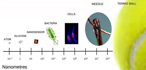
Scientists at the CSIC and at the IRB Barcelona have obtained a nanosensor based on small DNA fragments, using an advanced technique known as DNA origami. It has the size of 100 nanometres: it is a thousand times smaller than a bacteria and it would be necessary millions of sensors like this to fill the hole in a sewing needle.
The new molecular sensor can detect the activity of the human enzyme hAGT. This is interesting for anticancer research, as this enzyme is one of the targets for new drugs and it is one of the markers that can predict the success of a new treatment. It is, also, a substantial step in the control of DNA as building material for biomedical devices at the nanometric scale.
The work, published in the magazine Angewandte Chemie, has been developed by the group of Nucleic Acids Chemistry at the Institut de Química Avançada de Catalunya (IQAC-CSIC). The main author is Carmen Fàbrega, who is working in the same group but assigned to the IRB Barcelona.
Folding and stapling DNA as if it was paper
In the DNA origami technique -named after the traditional oriental art of folding paper to obtain figures- a long chain of DNA is naturally folded with small DNA segments (oligonucleotides) which work as staples, until the wanted structure is obtained.
The structure, which can have different shapes, can be used as a template to place proteins, nanoparticles, enzymes, nanowires or any other functional molecule following a predetermined pattern. The DNA has other advantages: it is rigid at the nanometric scale, it has an extraordinarily small size and an almost infinite possibility of combinations.
“In our work”, explains Ramon Eritja, leader of the Nuclei Acids Chemistry group, “we have used a single strand of viral DNA and 250 oligonucleotides ‘staples’ with which we did fold the DNA until we got a flat structure, with a rectangular shape”.
How the sensor works
Once the structure was created, scientists added to it two lines with sequences of DNA. One line contained an aptamer, a DNA single stranded nucleic acid, which has the capacity to recognize and bind to thrombin, a protein involved in blood coagulation. The other line had the same aptamer but with the genetic sequence “damaged”, as scientists modified it to make the aptamer unable to bind to thrombin.
When the samples –DNA similar to human DNA after a chemotherapy treatment- are placed on the nanosensor, it is possible to observe through Atomic Force Microcopy (AFM) if thrombin binds only to one line or to both of them (the one with the correct aptamer and the one with the ‘damaged’ aptamer). If thrombin binds to the ‘damaged’ aptamere means that the aptamer has been repaired by the hAGT enzyme contained in the sample.
Anticancer research
What is the hAGT enzyme? As Carmen Fàbrega explains, the hAGT is one of the so called repair kit enzymes of our cellular DNA. “Chemotherapy treatments are focussed on harming the DNA of cancerous cells. But, for some reason, cancer cells overexpress this hAGT enzyme, what makes them especially good to repair their own DNA after chemotherapy treatment”. This is one of the reasons that explains the emergence of resistance to cancer drugs.
Therefore, one of the strategies for new therapies is the search for hAGT inhibitors. Nevertheless “it is necessary to have new tools to view the function of hAGT in laboratory”, points out Fàbrega. It is very hard to do it with the current techniques, based on radioactivity essays. In this regard, the new biosensor is an alternative and reliable tool to detect the activity of this enzyme in a simplified way. Their use, nevertheless, is limited to basic research because of the expensive cost of AFM microscopy. “Maybe in the near future, the cost of AFM microscopy will decrease and this nanosensor could be used as a clinical tool, for molecular diagnosis”.
Reference article:
Origami as a DNA Repair Nanosensor at the Single-Molecule Level
Tintoré, M., Gállego, I., Manning, B., Eritja, R. and Fàbrega, C.
Angew. Chem. Int. Ed. (2013) doi: 10.1002/anie.201301293
About IRB Barcelona
The Institute for Research in Biomedicine (IRB Barcelona) pursues a society free of disease. To this end, it conducts multidisciplinary research of excellence to cure cancer and other diseases linked to ageing. It establishes technology transfer agreements with the pharmaceutical industry and major hospitals to bring research results closer to society, and organises a range of science outreach activities to engage the public in an open dialogue. IRB Barcelona is an international centre that hosts 400 researchers and more than 30 nationalities. Recognised as a Severo Ochoa Centre of Excellence since 2011, IRB Barcelona is a CERCA centre and member of the Barcelona Institute of Science and Technology (BIST).