
Organitzat per l'IRB Barcelona en col·laboració amb la Fundació Catalunya La Pedrera.
Presentation
Objectius
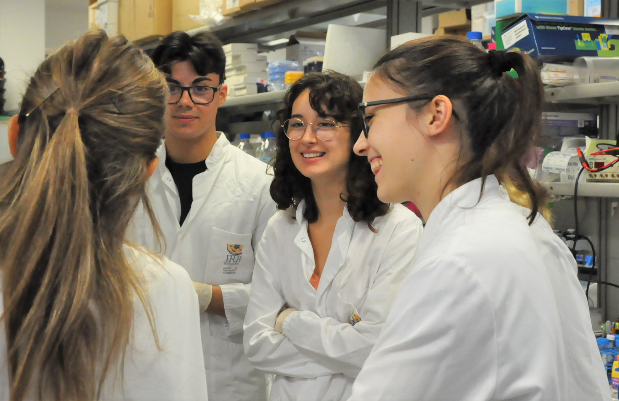
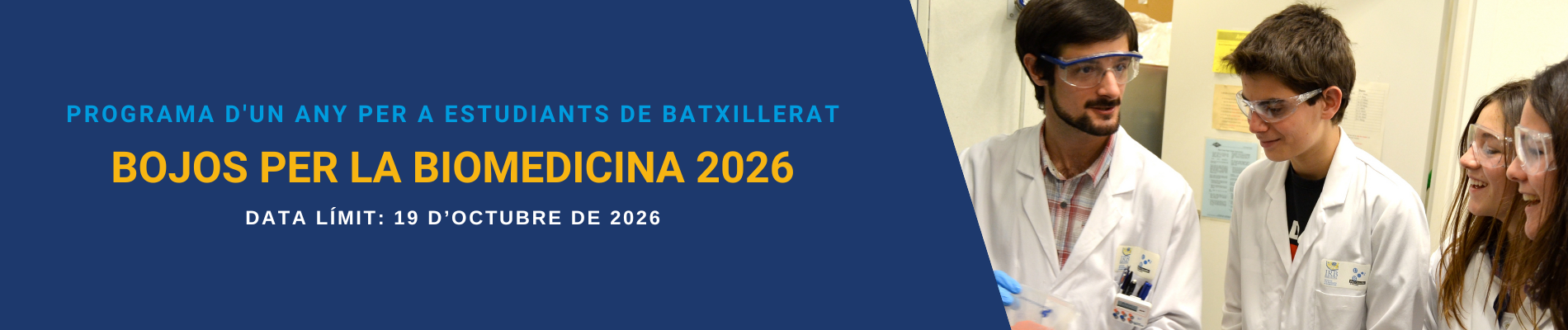
El Bojos per la Biomedicina és un curs dirigit als estudiants del primer any de batxillerat que desitgin explorar alguns dels descobriments fascinants que s'estan fent actualment en les ciències de la vida. A través d'aquest curs, els estudiants tindran l'oportunitat d'aprofundir el seu coneixement de la teoria i tècniques científiques en el camp de la biomedicina. Treballaran juntament amb investigadors joves per experimentar com es fa ciència en un institut de recerca internacional, guanyar una mica d'experiència pràctica en les últimes metodologies d'avantguarda i posicionar-se per a una possible carrera professional en les ciències de la vida.
Descripció del curs
Taller d'un any de durada sobre les ciències de la vida per a estudiants de batxillerat. Organitzat per l'IRB Barcelona dins el Programa "Bojos per la Ciència" de la Fundació Catalunya La Pedrera. "Bojos per la Ciència" de la Fundació Catalunya La Pedrera.
Aquest curs combina sessions teòriques i activitats experimentals pràctiques, que es duran a terme durant 16 dissabtes de l'any. El curs tractarà 10 temes científics actuals, que van des de la biologia cel·lular i molecular fins a la biologia estructural i computacional i la química, presentats per investigadors joves de l'IRB Barcelona. Els tres primers dissabtes es dedicaran a aquestes sessions teòriques generals per a tots els participants. Durant els deu dissabtes següents, es formaran grups de 5 estudiants que entraran als laboratoris per a les sessions pràctiques. Els estudiants participants s'hauran de comprometre a assistir al curs durant tot l'any.
Els estudiants rebran un certificat de participació en finalitzar el curs en una cerimònia especial de cloenda, a la que podran assistir pares i professors. A més, oferim la posibilitat de fer la part pràctica del Treball de Recerca de Batxillerat en els nostres laboratoris sota la tutela d'uns dels nostres investigadors o investigadores.
Idioma del curs
Totes les xerrades i sessions pràctiques es faran en anglès.

Dates i horaris
El curs tindrà lloc de gener a juny de 2026, els dissabtes de 10.00 a 14.00h.
- Dissabte 10 de gener: Inauguració oficial del curs i Xerrades 1-2-3
- Dissabte 24 de gener: Xerrades 4-5-6
- Dissabte 7 de febrer: Xerrades 7-8-9-10
- Dissabte 21 de febrer: Sessió pràctica 1
- Dissabte 28 de febrer: Sessió pràctica 2
- Dissabte 14 de març: Sessió pràctica 3
- Dissabte 21 de març: Sessió pràctica 4
- Dissabte 11 d'abril: Sessió pràctica 5
- Dissabte 18 d'abril: Sessió pràctica 6
- Dissabte 25 d'abril: Sessió pràctica 7
- Dissabte 9 de maig: Sessió pràctica 8
- Dissabte 16 de maig: Sessió pràctica 9
- Dissabte 6 de juny: Sessió pràctica 10
Preu del curs
Els participants hauran d'abonar a la Fundació Catalunya La Pedrera la quantitat de 425 euros. Consulteu l'oferta de beques al seu lloc web.
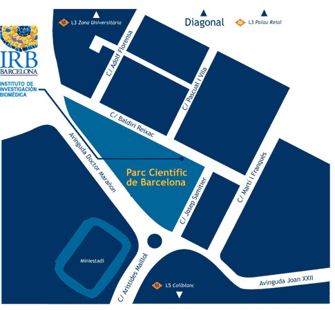
Lloc on es realitzarà el curs
Institut de Recerca Biomèdica (IRB Barcelona)
C/ Baldiri Reixac, 10
08028 Barcelona
Qui pot sol·licitar una plaça
Aquest curs està dirigit als estudiants de primer any de batxillerat que tinguin un interès i talent especials en els camps relacionats amb les ciències de la vida.
Els estudiants poden sol·licitar plaça a un màxim de 3 dels programes de la sèrie "Bojos per la Ciència" i finalment només podran participar en un d'ells.
Com sol·licitar una plaça
Les inscripcions s'hauran de realitzar aquí a partir del 15 de setembre 2025.
ls estudiants interessats hauran d'emplenar el formulari de sol·licitud i incloure una carta de motivació. També es demanarà una carta de recomanació directament de dos dels seus professors que coneguin bé l'alumne/a. En el cas de que l'estudiant hagi canviat de centre aquest curs, suggerim que sol·licitin les cartes als antics professors.
La data límit d'inscripció és el 19 d'octubre 2025 (23:59h).
El curs està obert a un total de 25 estudiants. Se seleccionaran els candidats en funció del seu expedient acadèmic, de les recomanacions dels seus professors i de la seva motivació per participar-hi. Es convidarà els candidats preseleccionats a fer entrevistes amb els organitzadors científics al novembre, després de les quals es farà la selecció final. El 25 de novmebre es comunicarà el resultat als estudiants. Es demanarà als estudiants seleccionats per participar-hi i als seus pares/tutors legals que signin una carta de compromís d'assistir a totes les sessions.
Col·laboradors
Per a qualsevol dubte, si us plau contacteu-nos a: irb_outreach@irbbarcelona.org
Dates importants
- 19 octubre 2025: Data límit d'inscripció
- 10 novembre 2025: Contacte amb els candidats preseleccionats
- 11 novembre - 23 novembre 2025: Entrevistes
- 24 novembre 2025: Contacte amb els candidats seleccionats
- 9 gener 2026: Inauguració oficial del curs
- 10 gener 2026: Inici del curs
- Acte de Clausura - data exacta a concretar
Programme
1. Destroy to discover the cure – Investigating Angelman syndrome
Karen Sfyaki (Targeted Protein Degradation and Drug Discovery Laboratory)

Angelman syndrome is a complex genetic disorder affecting primarily the brain. The main characteristics of this syndrome are delayed development, speech impairment, and problems with movement and balance and, in severe cases, epileptic seizures, and sleep disorders. Currently, there is no cure for Angelman syndrome, but we know that it is caused by the loss of a protein called UBE3A. This protein belongs to the family of E3 ubiquitin ligases. These enzymes mark proteins for degradation through the proteasome. The proteasome is the cell’s trash machinery that breaks down the proteins that are damaged or that are no longer needed. Therefore, without UBE3A, its substrates cannot be degraded by the proteasome and it will accumulate. However, the substrates of UBE3A are not yet known and they could be potential therapeutic targets for Angelman syndrome.
In recent years, a new application of the proteasome system has been developed to study proteins. Scientists have strategically managed to use the proteasome to their advantage by tricking it into degrading proteins that they are interested in. This approach is known as “targeted protein degradation”. To achieve this, we add a degradation tag to the protein of interest. This tag can be recognised by drugs called degraders, which bring the target protein and an E3 ligase closely together so that the protein can be marked for proteasomal degradation. This technology allows us to destroy any protein of interest and discover its downstream targets.
In this course, students will add the degradation tag to UBE3A using molecular cloning techniques and will become familiar with the techniques of DNA quantification, cloning and bacterial transformation.
2. Chemistry in a Click!: obtaining complex molecules through easy reactions
Ines Maria Garcia (Research Unit on Asymmetric Synthesis)

Chemists have always wanted to harness the power of molecular assembly to build complex molecules and obtain the widest range of applications. The logic behind this interest is simple—we are always in need of new molecular properties. These properties can be achieved by joining small molecular building blocks together—a process that becomes more difficult as the complexity of the molecules increases.
In 2001, Barry Sharpless introduced the concept of Click Chemistry as a new approach to synthesise complex drug-like molecules by using a few practical and reliable reactions. Click Chemistry includes chemical reactions that are performed with high selectivity, high yield, and a fast reaction rate, so that the “click” refers to the convenience afforded by snapping two objects together, like a luggage strap connection.
Click Chemistry has the following advantages over other types of reactions: it occurs in one pot; it is not disturbed by water; it generates minimal and usually harmless byproducts; and it has a high reaction specificity. Given these properties, this type of chemistry has been highly successful in recent years, leading its authors to win the Nobel Prize in Chemistry in 2022.
In this course, students will learn the basics of Click Chemistry and how to prepare these reactions. They will control these reactions using thin layer chromatography, detect the presence of both the reagents and the reaction products, and then finally purify the desired product.
3. Dealing with Stress – Learning survival mechanisms from the simplest eukaryotic organism
Ana Contreras, Ariadna Torres & Anna Granero (Cell Signalling Laboratory)


A fundamental property of living cells is the ability to sense and respond to fluctuations in their environment. Cells have developed a large number of signal transduction pathways that serve to adapt to these changes. In our lab, we aim to unravel how cells detect, respond, and adapt to environmental variations.
Working with the budding yeast Saccharomyces cerevisiae, a well-established eukaryotic model organism that has the following advantages: ease of manipulation; short life span; ability to produce a large number of offspring; and a sequenced genome. We use S. cerevisiae to study the adaptive eukaryotic responses to a variety of environmental stresses. Remarkably, many processes initially discovered and studied in yeast, such as transcription regulation, stress-signalling transduction, and cell cycle regulation are conserved in higher eukaryotes.
Yeast and mammals share a conserved family of proteins known as Stress-Activated Protein Kinases (SAPKs) that sense and respond to environmental changes. The activation of SAPKs leads to a set of adaptive responses that involve the modulation of several physiological processes, including gene transcription, cell metabolism, protein translation, and cell cycle progression.
In this course, students will learn how to manipulate and work with S. cerevisiae to understand how cells detect and respond to external signals in order to ensure survival. For this purpose, we will study the activation of Hog1 SAPK upon different stress conditions and examine the cell fitness of various mutant strains when subjected to environmental insults.
4. Using the protein-eating machine to treat cancer
Jake Antony Ward (Targeted Protein Degradation and Drug Discovery Laboratory)

Control over the concentration, structure, and location of proteins is important for the health and fitness of our cells. When proteins are damaged or are no longer needed, they are broken down within the cell in a process known as degradation. One of the main cellular systems involved in degrading unwanted or damaged proteins is called the proteasome. The proteasome is made up of many enzymes, such as proteases, which recognise and break down these proteins. For a damaged or unwanted protein to be recognised and broken down by the proteasome, a specific label, known as ubiquitin, is usually added to the protein. Certain enzymes known as ubiquitin ligases are involved in this tagging process.
In recent years, drugs have been developed that use this proteasome system in our cells to degrade certain proteins. This is an exciting new strategy, as it can allow us to remove a given protein from a tumorogenic cell. This approach is known as Targeted Protein Degradation (TPD). This technology works by using a drug that brings the ubiquitin ligase and the target protein for degradation closer together. The protein is thus labelled with the ubiquitin-tag and is recognised by the proteasome and destroyed.
In this course, students will focus on a type of cancer known as Gastrointestinal Stromal Tumour (GIST). This cancer typically develops in the muscle cells of the gut. When mutated, the KIT protein is the molecule responsible for the formation and growth of this type of cancer. We are interested in designing drugs using the TPD strategy to remove KIT from GIST cancers. This line of research is exciting as it may lead to new treatments for GIST. Students will gain hands-on experience with the key techniques of gel electrophoresis and Western blotting to monitor the degradation of KIT in human cancer cells.
5. Machine learning to fight drug resistance
Elena Pareja (Structural Bioinformatics and Network Biology Laboratory)

Drug resistance, which occurs when previously vulnerable cells become unresponsive to specific drugs, poses a significant public health concern worldwide. Antimicrobial resistance (AMR) exemplifies this issue, with estimates indicating that the annual death toll resulting from AMR could skyrocket from 700,000 to 10 million people by 2050. Similarly, drug resistance affects the efficacy of cancer treatment, as patients may develop resistance following a relapse. The primary driver of drug resistance involves mutations in the DNA sequence of proteins associated with the drug's mode of action.
To comprehensively study this phenomenon, a crucial step involves analysing DNA sequencing data and establishing connections with protein structure and function to enhance our understanding of resistance mechanisms and devise strategies to overcome them. Machine learning has emerged as a valuable tool in predicting drug resistance by leveraging sequencing data.
In this course, students will have the opportunity to explore public databases and extract relevant biological information concerning drug resistance. They will work hands-on with sequencing data to understand the significance of mutations and observe how alterations in the sequence impact the shape and function of proteins. Finally, they will build a small machine-learning model to predict drug resistance on the basis of sequence data.
6. DNA Detectives: Cracking the Mouse Genome Code
Elsa Tusquets (Translational Control of Cell Cycle and Differentiation)

Ever wondered how scientists study human diseases or develop new medicines? A lot of the time, they do it by studying genetically modified mice! These mice are created with specific genetic changes to help us understand everything from cancer to diabetes. But how do we know which mouse has which genetic change?
Mice are an ideal model for studying human diseases due to their high degree of genetic and physiological similarity to humans. This genetic homology allows scientists to create genetically modified mice that mimic human conditions, providing a controlled environment to study disease progression and test new therapies. Scientists can introduce specific genetic mutations into mice to study how these mutations cause disease. For example, a "knockout" mouse is created by deactivating a specific gene. If this gene is known to be involved in a human disease, the knockout mouse can be used to study the disease's mechanisms and test new drugs.
In this experimental session, you will learn the fundamental molecular biology techniques used to identify these genetic modifications. You'll perform Polymerase Chain Reaction (PCR) to amplify specific DNA sequences from different mice, then use agarose gel electrophoresis to separate these DNA fragments by size. By analysing the resulting band patterns you’ll effectively determine the genotype of each mouse.
7. The Dance of Chromosomes: unveiling the hidden moves of cell division
Javier Manzano (Translational Control of Cell Cycle and Differentiation)

Every day, our cells divide to keep us growing, healing, and alive. In this process, called mitosis, chromosomes are carefully copied and then evenly shared so each new cell gets exactly what it needs. But when this choreography goes wrong, cells may end up with too many or too few chromosomes—a mistake known as aneuploidy. This imbalance in chromosome number creates genetic instability, which can push cells toward uncontrolled growth and is a driving force behind many types of cancer. With fluorescence microscopy, scientists can make this hidden dance visible, watching chromosomes move in real time and spotting the errors that sometimes occur. In this course, students will step into the scientist’s role: prepare samples, apply fluorescent stains, and use advanced microscopy to observe chromosome segregation—and discover what happens when the dance goes wrong.
8. 3D genome organization: from protein disorder to gene control in cancer
Magalí Scocozza (Laboratory of Molecular Biophysics)

A typical concept is that proteins adopt a native structure encoded by their amino acid sequence. However, some proteins contain long stretches called intrinsically disordered regions (IDRs) that do not fold into a fixed conformation, and these constitute nearly 40% of the human proteome. Instead, they remain flexible, explore different conformations, and can sometimes drive the formation of separate phases known as condensates. A human chromosome has millions of base pairs and could, in principle, form an astronomical number of 3D arrangements. Yet in vivo, chromosomes fold into reproducible higher-order structures. Chromatin fibers—DNA wrapped around histone proteins, like beads on a string—do not adopt a single rigid state, nor are they completely unstructured. Instead, they are organized by multiple mechanisms that bias them toward certain conformations within a diverse ensemble. Thus, both IDRs and chromatin can explore multiple conformational states, but depending on the guiding forces present, they can be switched into particular ensembles that carry out specific functions. In this way, transcription factors and other chromatin-binding proteins can actively shape and interact with chromatin states to regulate gene expression and alter cell fate.
In this workshop, students will examine immunofluorescence images of human prostate cancer cells, where the androgen receptor (AR) forms nuclear condensates after addition of dihydrotestosterone. Here they will learn that while some transcription factors can remodel chromatin, AR does not change chromatin states itself but selects among them, binding to accessible regions to regulate gene expression. Through this activity, students will explore how physical forces and protein disorder influence genome organization, gene regulation, cell fate decisions, and ultimately disease.
9. Mirror, mirror on the wall… Which is the most helpful insect of them all? – Getting to understand tumorigenesis through the fly
Berta Muñoz, Carlos Escribano (Development and Growth Control Laboratory)


The fruit fly Drosophila melanogaster has been widely used as a model organism for more than one hundred years to address biological questions in various fields. This organism has emerged as a potent tool for genetic manipulation, offering innumerable possibilities to analyse the detailed interaction between cells and tissues.
One question could be raised: how can a tiny organism like D. melanogaster be used to understand the pathways of a disease as complex as cancer? The answer is quite simple. Many biological mechanisms are well-conserved across evolution, including those related to pathological conditions. This conservation allows scientists to translate the knowledge acquired from less complex organisms to more complex ones. For instance, 75% of the genes related to human diseases and 68% of the genes related to human cancer have a counterpart in the fly. We are more similar to the fly
than we may think!
In this course, students will learn how to study the complex process of tumorigenesis using this model organism, both with a local and systemic approach. We will focus mainly on carcinomas, the most common type of tumour diagnosed in humans.
Carcinomas are derived from epithelial tissue, such as the skin, and they can become invasive or metastatic by spreading beyond the primary tissue layer and surrounding tissues or organs. In aggressive cancer cells, this transition is mediated by the activation of the EMT (Epithelial to Mesenchymal Transition) program, which causes the cells to undergo morphogenetic alterations that increase invasive capacity.
In this course, students will be introduced to the first steps in fly genetics. We will cross flies, identify genetic markers and balancer chromosomes, and apply other genetic tools we use in the lab every day to generate tumorigenic flies and study them. Students will also learn about the anatomy of the fly in adult and larvae stages and use advanced microscopy to detect several genetic markers in the tumoral
tissues. Finally, they will perform in vivo dissections and the respective immunohistochemistry.
10. Mitochondria: more than the powerhouse of the cell
Jake Pickering, Valeria Lupi (Miochondrial Biology and Tissue Regeneration)


Mitochondria are not only the ‘powerhouse of the cell’ but are heavily involved in sending signals that control how cells behave. As well as being maternally inherited, not all mitochondria are made equal. Individuals from different parts of the world have varying small nucleotide polymorphisms with the mitochondrial DNA (mtDNA), that result in different mutations in the 37 genes encoded by the mtDNA; referred to as mitochondrial haplogroups. Such variances dictate a cells metabolic preference, whilst increasing the risk and severity of disease pathologies. This includes systemic inflammation, cardiac dysfunction and cancer progression.
Lymphocytes with compromised mitochondrial function demonstrate pro-inflammatory behaviour and a reduced ability for T cells to fight infection or disease. Protecting the functionality of the mitochondria in these cells has been shown to not only enhance their metabolism but has also been shown to reduce inflammation in organs. This is crucial for slowing disease development. However, skin cancers such as melanoma can exploit the metabolic preferences of these haplogroup variances, leading to the development of metastasis, and the suppression of immune cells in the tumour microenvironment.
Within this course, students will utilise Flow cytometry to assess the metabolic preferences of tumour cells. They will assess this on melanoma cells derived from cells with the same nuclear genotypes but differing mtDNA.
Venue
Crazy About Biomedicine course will take place at the IRB Barcelona facilities.
Institute for Research in Biomedicine (IRB Barcelona)
Parc Cientific de Barcelona
C/ Baldiri Reixac, 10
08028 Barcelona