
Organized by IRB Barcelona in collaboration with the Fundació Catalunya La Pedrera.
Presentation
Objectives
Crazy About Biomedicine is a course directed at students in their first year of baccalaureate who wish to explore some of the discoveries being made in biomedicine. Through this course, students will have a chance to deepen their knowledge of scientific theory and techniques in the field of biomedicine. They will work alongside our young researchers to get a taste for what doing science in a top international research institute is like, gain some experience in cutting-edge methodologies, and position themselves for a potential career in the life sciences.
Course Description
6 month workshop in the life sciences for high-school students organized by IRB Barcelona within the series "Crazy About Science" of the Fundació Catalunya La Pedrera.
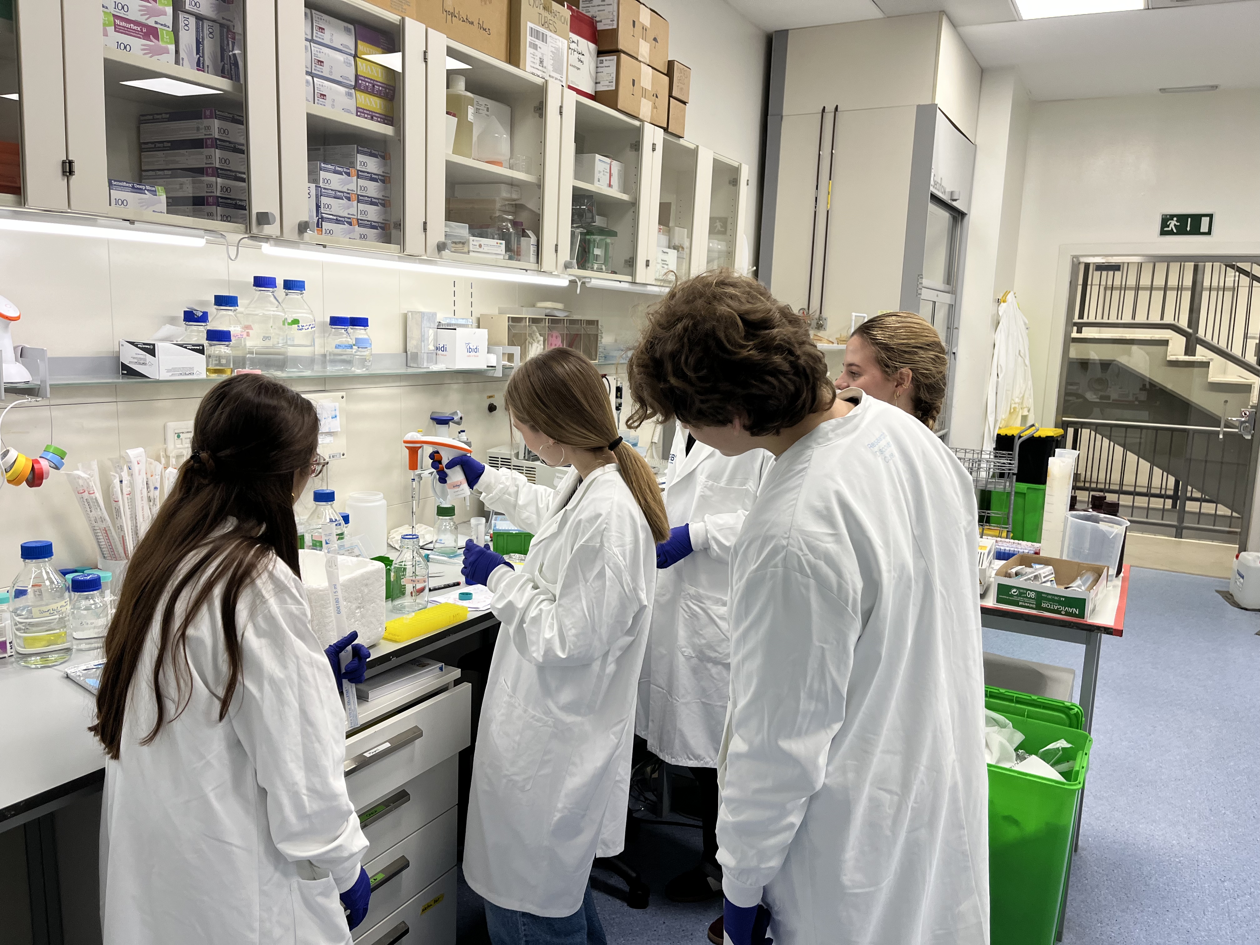
This course includes a combination of theoretical lectures and practical hands-on experimental activities, which take place on 13 Saturdays throughout the year. Presented by IRB Barcelona PhD students and postdocs, the course will cover 10 hot scientific topics, ranging from cell and molecular biology to structural and computational biology, and chemistry. The first 3 Saturdays will be dedicated to doctorand theoretical sessions for all participants. Over the next 10 Saturdays, groups of 5 students will enter the laboratories for practical sessions. Participants must commit themselves to attend the whole course.
Students will receive a Certificate of Participation upon successful completion of the course at a special ceremony to which parents and teachers will be invited. Moreover, we offer the possibility of performing the practical part of the Treball de Recerca of Batxillerat in our laboratories under the supervision of one of our researchers.
Course language
All lectures and practical sessions will be conducted in English.

Course dates and times
The course will run from January to June 2027, Saturdays 10.00-14.00h.
- Sat. 9 January: Official course inauguration and Talks 1-2-3
- Sat. 23 January: Talks 4-5-6-7
- Sat. 6 February: Talks 8-9-10
- Sat. 20 February: Practical session 1
- Sat. 27 February: Practical session 2
- Sat. 6 March: Break
- Sat. 13 March: Practical session 3
- Sat. 20 March: Practical session 4
- Sat. 27 March: Easter Saturday (holiday)
- Sat. 3 April: Practical session 5
- Sat. 10 April: Practical session 6 or Open Day
- Sat. 17 April: Practical session 7
- Sat. 24 April: Practical session 8
- Sat. 1 May: Labour Day (holiday)
- Sat. 8 May: Practical session 9 or Open Day
- Sat. 15 May: Practical session 10
- Sat. 22 May: Remaining practical session of the ODD
- Sat. 29 May: Scientific Communication Workshop
- Sat. 5 June: Break
- Sat. 12 June: Closing session and families' day
Course fees
There is a fee of 425 euros payable directly to the Fundació Catalunya La Pedrera. Take a look at the scholarships available at their webpage.
Course location
Institute for Research in Biomedicine (IRB Barcelona)
C/ Baldiri Reixac, 10
08028 Barcelona
Who can apply
This course is directed toward students in the first year of their baccalaureate (only), who have a special interest and talent in the fields related to the life sciences (primarily biology and chemistry).
Students may apply to a maximum of 3 programmes within the "Crazy About Science" series, and can participate in only one.
How to apply
Applications have to be submitted from here, starting on 14 September.
Interested students must fill in the online application form and include a letter of motivation. A letter of recommendation will be requested directly from two of their teachers, who should know the applicant well. If the applicant has recently changed school then the letters of recommendation should be requested from his/her former teachers.
The deadline for registration is 18 October 2026 (23:59h).
The course is open to a total of 25 students. Candidates will be selected on the basis of their academic record, teacher recommendations and motivation to participate. A shortlist of candidates will be invited for interviews with the scientific organisers in November after which the final selection will be made. Students will be informed of the outcome by 23 November. The students selected to participate and their parents/guardians will be asked to sign a letter of commitment to attend all sessions.
In collaboration with
If you have any question, please contact us at irb_outreach@irbbarcelona.org
Important dates
- 18 October 2026: Registration deadline
- 9 November 2026: Contact with preselected candidates
- 10 November - 22 November 2026: Interviews
- 23 November 2026: Contact with selected candidates
- 8 January 2027: Official course inauguration
- 9 January 2027: Course start
- 4 June 2027: Closing ceremony - exact date to be confirmed
Programme
1. DNA Detectives: Cracking the Mouse Genome Code
Andrea Zingaro (Translational Control of Cell Cycle and Differentiation)

Ever wondered how scientists study human diseases or develop new medicines? A lot of the time, they do it by studying genetically modified mice! These mice are created with specific genetic changes to help us understand everything from cancer to diabetes. But how do we know which mouse has which genetic change?
Mice are an ideal model for studying human diseases due to their high degree of genetic and physiological similarity to humans. This genetic homology allows scientists to create genetically modified mice that mimic human conditions, providing a controlled environment to study disease progression and test new therapies. Scientists can introduce specific genetic mutations into mice to study how these mutations cause disease. For example, a "knockout" mouse is created by deactivating a specific gene. If this gene is known to be involved in a human disease, the knockout mouse can be used to study the disease's mechanisms and test new drugs.
In this experimental session, you will learn the fundamental molecular biology techniques used to identify these genetic modifications. You'll perform Polymerase Chain Reaction (PCR) to amplify specific DNA sequences from different mice, then use agarose gel electrophoresis to separate these DNA fragments by size. By analysing the resulting band patterns you’ll effectively determine the genotype of each mouse.cular cloning techniques and will become familiar with the techniques of DNA quantification, cloning and bacterial transformation.
2. Machine learning to fight drug resistance
Aurora Desideri & Dylan Dalton (Structural BiologyStructural Bioinformatics and Network Biology)
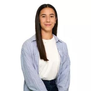
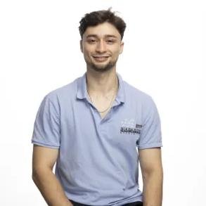
Drug resistance, which occurs when previously vulnerable cells become unresponsive to specific drugs, poses a significant public health concern worldwide. Antimicrobial resistance (AMRemplifies this issue, with estimates indicating that the annual death toll resulting from AMR could skyrocket from 700,000 to 10 million people by 2050. Similarly, drug resistance affects the efficacy of cancer treatment, as patients may develop resistance following a relapse. The primary driver of drug resistance involves mutations in the DNA sequence of proteins associated with the drug's mode of action. To comprehensively study this phenomenon, a crucial step involves analysing DNA sequencing data and establishing connections with protein structure and function to enhance our understanding of resistance mechanisms and devise strategies to overcome them. Machine learning has emerged as a valuable tool in predicting drug resistance by leveraging sequencing data. In this course, students will have the opportunity to explore public databases and extract relevant biological information concerning drug resistance. They will work hands-on with sequencing data to understand the significance of mutations and observe how alterations in the sequence impact the shape and function of proteins. Finally, they will build a small machine-learning model to predict drug resistance on the basis of sequence data.
3. Mirror, mirror on the wall… Which is the most helpful insect of them all? – Getting to understand tumorigenesis through the fly
Carlos Escribano & Nikita Nikita (Development and Growth Control Laboratory)
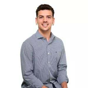

The fruit fly, Drosophila melanogaster, has been widely used as a model organism for more than one hundred years to address biological questions in various fields. This organism has emerged as a potent tool for genetic manipulation, offering innumerable possibilities to analyse the detailed interaction between cells and tissues.
One question could be raised: how can a tiny organism like D. melanogaster be used to understand the pathways of a disease as complex as cancer? The answer is quite simple. Many biological mechanisms are well-conserved across evolution, including those related to pathological conditions. This conservation allows scientists to translate the knowledge acquired from less complex organisms to more complex ones. For instance, 75% of the genes related to human diseases and 68% of the genes related to human cancer have a counterpart in the fly. We are more similar to the fly than we may think! In this course, students will learn how to study the complex process of tumorigenesis using this model organism, both with a local and systemic approach. We will focus mainly on carcinomas; the most common type of tumour diagnosed in humans.
Carcinomas are derived from epithelial tissue, such as the skin, and they can become invasive or metastatic by spreading beyond the primary tissue layer and surrounding tissues or organs. In aggressive cancer cells, this transition is mediated by the activation of the EMT (Epithelial to Mesenchymal Transition) program, which causes the cells to undergo morphogenetic alterations that increase invasive capacity.
In this course, students will be introduced to the first steps in fly genetics. We will cross flies, identify genetic markers and balancer chromosomes, and apply other genetic tools we use in the lab every day to generate tumorigenic flies and study them. Students will also learn about the anatomy of the fly in adult and larvae stages and use advanced microscopy to detect several genetic markers in the tumoral tissues. Finally, they will perform in vivo dissections and the respective immunohistochemistry.
4. Nanometric Changes with Macroscopic Consequences: Protein Structure, Function, and Disease
Jana Bagan (Structural Characterization of Macromolecular Assemblies)

Proteins are the machinery of the cell: from digesting food to copying DNA, every task in a living organism is carried out by proteins. They are synthesized in the ribosome as a polypeptide chain that folds into a precise 3D shape upon exposure to the aqueous intracellular environment. The 3D structure of proteins determines their function, and even small shifts in their architecture can completely shut down their activity or alter their role in the cell, leading to disease. Structural biology gives us the power to map these exact atomic flaws, allowing scientists to design targeted drugs that restore normal protein function or block harmful cellular activity. Traditionally, the structure of proteins has been studied through X-ray crystallography, where protein crystals are subjected to an X-ray beam to obtain diffraction patterns from which the 3D positions of individual atoms can be determined.
In this workshop, students will explore the structure-function relationship of proteins by assessing how environmental stress conditions alter enzyme performance. They will get a taste of protein crystallization and learn how X-ray crystallography generates 3D atomic maps. Finally, we will inspect the structures of our study proteins using 3D molecular visualization software (PyMOL, ChimeraX) to match our experimental findings with structural data, identifying which atomic features are responsible for protein function and how they are impaired under stress.
5. From Mouse to Microscope: Uncovering the Secrets of Tissues
Maria Díaz (Histopathology Facility)
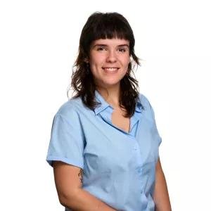
How can scientists understand what happens inside an organism when a disease develops or when a new treatment is tested? One way is by studying tissues under the microscope. In biomedical research, mice can be used as experimental models to investigate biological processes and diseases. By examining their tissues, scientists can detect changes that cannot be seen with the naked eye and gain a better understanding of what is happening at the cellular level.
Histopathology is the study of tissues and their microscopic structure, and it plays an important role in biomedical research. Researchers can compare tissues from different experimental conditions to investigate how a disease progresses or how a treatment affects an organ. To do this, tissues are carefully processed, embedded, cut into very thin sections and stained before being observed under a microscope. These techniques transform a small piece of tissue into a source of valuable scientific information.
In this experimental session, students will discover how researchers go from a mouse tissue sample to a microscopic image. They will learn how histological samples are prepared, observe different mouse tissues under the microscope and identify basic cellular and tissue structures. Finally, they will compare tissue samples from different experimental conditions and investigate the changes they can observe, just like scientists working in a real research laboratory.
6. Drug-tolerant persister cells: can we see the cancer cells that hide from treatment?
Maria Solivellas, Núria Gendrau & Joana Martins (Growth Control and Cancer Metastasis)
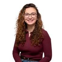

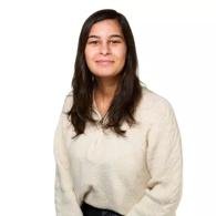
Breast cancer is the most common cancer among women. Although most patients have a good prognosis, around one in three will eventually experience a relapse. This happens when cancer cells escape from the primary tumor and travel to other organs, a process known as metastasis. Particularly intriguing is estrogen receptor-positive breast cancer (ER+ BCa), which can come back many years after the initial diagnosis, sometimes even 20 years later. How can cancer cells survive treatment for so long? One possible explanation is a population of drug tolerant persister (DTP) cells. While standard treatments eliminate rapidly growing cancer cells, some cells can temporarily enter a slow, inactive state, allowing them to survive treatment and remain hidden for a long time. Later, they can “wake up” and cause a metastatic relapse.
In our lab, we use a color-based reporter system based on the protein p27, which allows us to identify cells that slow down their activity in response to treatment. During the course, students will introduce the p27 reporter into breast cancer cells, treat them and observe their p27 patterns. Finally, they will analyze the results and connect them with data from BCa patients, discovering how laboratory experiments can help us understand cancer relapse.
7. Hunting for Drugs: How Do We Find New Treatments for Cancer?
Maria Urrutia (Stress Signaling in Cancer)
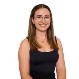
Developing a new drug is a long and complex process that begins with a simple question: how can we find a molecule that can change the behaviour of a disease? In cancer research, scientists search for vulnerabilities that can be exploited to selectively affect cancer cells while leaving healthy cells as unharmed as possible.
One way to discover these vulnerabilities is through drug screening, in which large numbers of compounds are tested to identify those that produce a desired biological effect. Scientists can also study specific genes and proteins to understand whether they are important for cancer cell survival. By combining these approaches, it is possible to discover new potential drug targets and identify compounds that could be developed into future treatments.
But finding a molecule that works is only the beginning. Once a promising compound, or hit, has been identified, scientists need to determine how strongly it acts, whether its effect can be reproduced, and how changes to its chemical structure affect its activity. This process, known as drug discovery and optimisation, involves concepts such as potency, IC50 and structure–activity relationships (SAR).
In this activity, students will explore how researchers search for new anticancer drugs and how genetic and chemical information can be combined to identify potential therapeutic vulnerabilities. They will analyse experimental samples to determine whether a gene has been successfully knocked out, interpret dose–response curves to compare the potency of different compounds, and examine chemical structures to identify relationships between molecular structure and biological activity. Through these experiments, students will follow the logic of a real drug-discovery project, from identifying a potential vulnerability to evaluating and improving a candidate compound.
8. Blood vessels: more than just pipes!
Marta Fidalgo (Growth Control and Cancer Metastasis)
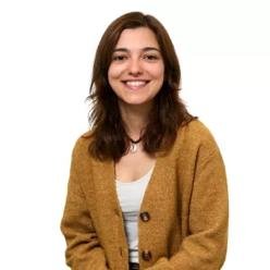
Blood vessels form a vast network that transports blood. They deliver oxygen and nutrients throughout our bodies and carry away waste. These functions are essential for life, but there is much more to blood vessels than transport! They are built from living cells that sense their surroundings, exchange signals with neighbouring cells and adapt to the needs of different tissues.
In this course, students will meet the endothelial cells that line blood vessels and discover why not all vessels are the same. We will explore how vascular networks grow and remodel to support healthy processes, such as adaptation to physical exercise, and how cancer can exploit blood vessel growth and communication to support tumour progression. Students will discover that blood vessels do more than respond to their environment: they can actively influence how other cells behave.
During the practical session, students will use microscopy to explore vascular structures and investigate endothelial cell behaviour through hands-on laboratory work. They will make predictions, compare experimental conditions and interpret their observations, connecting what they see under the microscope to the roles of blood vessels in health and disease.
By the end of the workshop, students will understand how scientists study blood vessels and why these dynamic, diverse structures are much more than just pipes!
9. Finding Cancer’s Weak Spots: How Can We Make Cancer Cells More Vulnerable?
Oriol Gómez (Stress Signaling in Cancer)
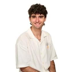
What makes a cancer cell different from a healthy cell? How can cancer survive treatments designed to kill it? In this workshop, you will discover how scientists search for the weak points of breast cancer cells and explore how these discoveries could contribute to better treatments for patients.
You will learn what cancer and chemotherapy resistance are, why some cancer cells can survive treatment, and which techniques scientists use to study these questions. You will also discover what it takes to become a scientist—from school and university to working in a real research laboratory—and learn how scientific research works, from generating data to drawing conclusions that can contribute to advances in medicine. In the practical part of the workshop, you will use two techniques routinely employed in cancer research laboratories—colony formation and CCK8 assays—to investigate how cancer cells grow and survive after treatment. We will also use interactive tools to make this workshop more hands-on, and interactive for everyone. So be ready to have fun!
The question is simple: where is cancer’s weak spot? Let’s find out.
10. DNA Fingerprinting: Solving a Genetic Mystery in the Lab
Giulia Viola (Microtubule organization in cell proliferation and differentiation)

How do we inherit our traits from our parents? How can scientists test who is related to whom using just a simple cheek swab? In this workshop, you will step into a real research lab to explore how DNA profiling works and how scientists analyze genetic markers to solve biological mysteries.
You will learn what DNA actually looks like, how traits are passed down through generations, and how techniques like PCR and gel electrophoresis allow us to read genetic profiles. You will also discover what it takes to become a scientist—from studying at university to working in a biomedical laboratory—and see how everyday research leads to real-world discoveries.
In the practical part of the workshop, you will isolate your own DNA from cheek cells and run an agarose gel electrophoresis experiment to analyze pre-prepared DNA samples and solve a parentage case. You will practice using micropipettes, loading gels, and inspecting glowing DNA bands under blue light to match the genetic profiles.
In science, data gives us the answer. Let’s run the gel, read the results, and make the final call.
Venue
Crazy About Biomedicine course will take place at the IRB Barcelona facilities.
Institute for Research in Biomedicine (IRB Barcelona)
Parc Cientific de Barcelona
C/ Baldiri Reixac, 10
08028 Barcelona